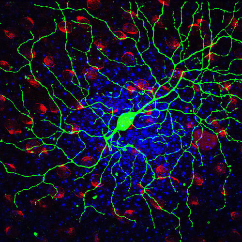
This is a GFP-tagged mouse retinal ganglion cell (in green), overlaid on a layer of cells with DAPI-stained nuclei (in blue) and ChAT-stained starburst amacrine cells (in red), taken with a tri-channel confocal fluorescence microscope.
I captured this image during a research project supervised by Sebastian Seung at the Massachusetts Institute of Technology and Richard Masland at Harvard. The goal of the collaboration was to develop an improved classification system for ganglion cells based upon their position in the retina and upon the morphology, or architecture, of their dendritic arbors (which behave like antennae).
I developed a series of semi-automated algorithms to digitally reconstruct the 3-D shape of the neuron from a stack of image slices and to calculate the position of the neuron's dendritic arbor in the retina. One issue that has plagued previous attempts to classify ganglion cells is that retinal tissue commonly becomes warped and wrinkled on the slide, which hinders precise localization of the arbor's depth.
Our approach uses the DAPI and ChAT fluorescence channels to mark known landmarks in the retina in the image slices. We can use these markers to unwarp the tissue digitally and thereby obtain improved estimates of each cell's stratification depth. By digitally unwarping the tissue and automating the reconstruction and localization procedure, the classification toolkit I developed this summer will allow ganglion cells to be classified faster and more accurately than was previously possible. With the help of this toolkit, researchers can begin to build a quantitatively accurate retinal "parts list."