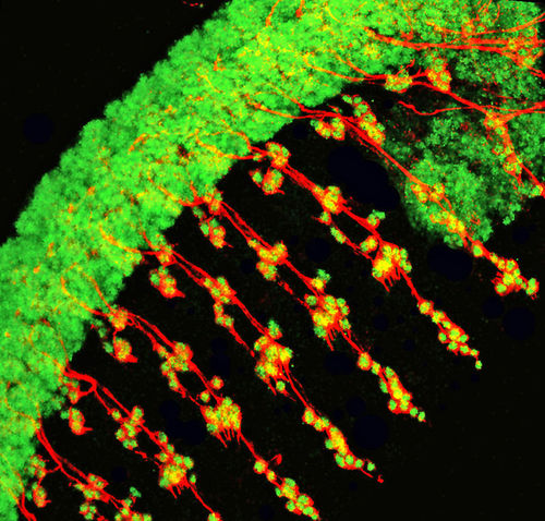
This image shows the organization of Drosophila melanogaster (fruitfly) embryonic neurons. Drosophila embryos have eight abdominal segments, where repeated patterns of peripheral neurons are observed. In order to study the patterns, I used fluorescent immunostaining to label different components of the neurons.
Here, red indicates the axons of neurons and green represents all nuclei in both the central nervous system and the peripheral nervous system. The thick bundle of green cells represents the ventral nerve cord (or central nervous system) which extends throughout the embryo and forms a future head structure at the anterior (upper right in the image).