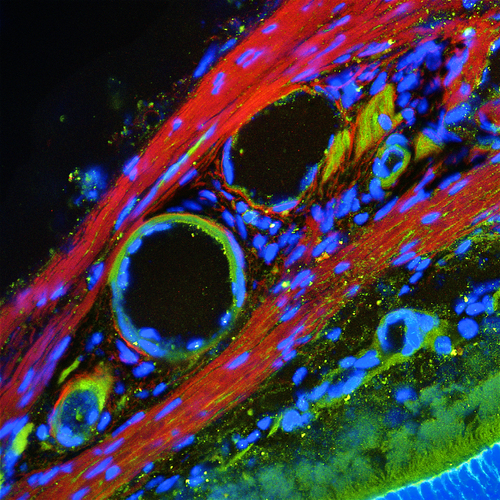
This image is a frozen section of a mouse eye that shows connective tissue, blood vessels, sclera (the white part of the eye), and part of the retina. The green parts of this image are the calcium-binding protein Calretinin, which have been stained with an antibody. Cell nuclei, stained with Hoescht 33258, are blue in this image.
Calcium-binding proteins participate in cell-signaling pathways by binding to calcium ions and thus power many aspects of a cell's functioning. The circular structures we see here are blood vessels that fuel the function of the eye by providing nutrients. A sliver of the retina -- the most energy-hungry part of the eye -- can be seen at the lower right, its turquoise hair-like photoreceptor outer segments fringing the adjacent bright-blue cell body layer.