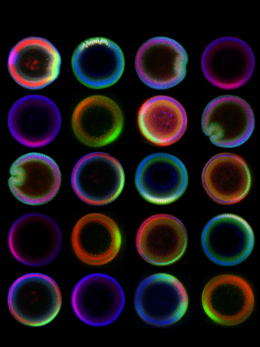
These images are vertical cross-sectional images of embryos of Drosophila melanogaster -- otherwise known as the common fruit fly. The images, obtained using a confocal microscope, are of embryos stained with antibodies in order to visualize molecules that subdivide the embryo into three tissue types: muscle, nervous system, and skin.
Obtaining such images is an engineering challenge since it requires upright positioning of a tiny embryo, which is ellipsoid in shape and only a half-millimeter long.
In collaboration with Lu lab at Georgia Tech, we have developed a microfluidic device to trap and orient a large number of embryos vertically. This technique can be used to quantify spatial profiles of signaling molecules, which can be used to develop mathematical models and eventually to understand the processes that drive the development of the embryo.