I am conducting a project where we look for viral and cellular proteins that change their sub cellular localization during human cytomegalovirus (HCMV) infection of human fibroblasts. We have generated a library of clonal hybridoma cell lines, screened the resulting antibodies with an immunofluorescence (IF) based high throughput assay and started to characterize the antibodies and their target proteins. We have screened approximately 35,000 hybridoma cell lines and from this we selected about 600 clones for further characterization.
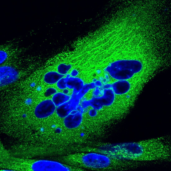
The picture Falling Apart is one of the first pictures out of the high throughput IF screen. It was part of the proof of principle that my work—taking 2½ years and hundreds of hours—was actually successful. I do not normally remember the feeling I have while taking pictures since I spend quite some time with our scope. But I actually remember this one—it was one of the most exiting days in my career, since I got the proof that a huge screen had worked. After my scope time that day I went straight to Triumph and had a couple of beers.
The picture shows a human fibroblast cell which is stained by immunofluorescence with an antibody derived from the screen described above (green). This antibody is directed against an unknown cellular protein which possibly plays a roll in HCMV infection. This protein is actually just now in our mass spectrometer facility for ultimate characterization. The cellular DNA (nucleus) is stained in blue. The cell probably is undergoing apoptosis since the nucleus is undergoing fragmentation ("falling apart").