
February 26, 2001
Vol. 90, No. 18
Big picture begins with smallest details
Steven Schultz
|
|
|
|
|
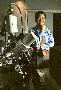
Nan Yao helps researchers enhance images --
and their work -- through resources like this new microscope in
the Princeton Material Institute's Imaging and Analysis Center.
|
He is the director of the Imaging and Analysis Center at the Princeton Materials Institute, a place where faculty members come to capture glimpses of everything from fruit fly cells to clusters of atoms in high-tech materials.
Located in the first floor of the Materials Institute's Bowen Hall, the imaging center consists of seven room-sized electron microscopes and computing labs for analyzing the images they produce. But it is more than just equipment. Yao, a physicist by training, is a master of the painstaking and creative process of harnessing a microscope's power to create clear images. Indeed, his contributions have made him an author or co-author of about 100 research papers from many labs.
"Nan is a top-notch electron microscopist," said Ilhan Aksay, a professor of chemical engineering who has co-written scores of papers with Yao. Likening the microscopes to a garage full of cars, Aksay said Yao is far more than just a mechanic. "You have the cars running, but you really don't know where to drive them. We work together on the interpretation of the images, and he really contributes intellectually."
The University established the Princeton Materials Institute in 1993 and recruited Yao that same year to help build the imaging capabilities that are required for materials research. After receiving a 1990 Ph.D. in condensed matter physics and electron microscopy from Arizona State University, Yao went to work in industry, first at the Shell Development Co., then at the Exxon Research and Engineering Co.
In coming to Princeton, Yao was attracted by the opportunity to build an imaging program where there was essentially none before. "We went from zero to one of the best imaging centers in the country," he said.
Powerful electron microscopes are a critical tool in materials science. "If you can't look at your material, you can't claim you are doing material science," said Aksay, who holds a joint appointment in the materials institute.
"We couldn't fulfill our mission without it," said institute director Anthony Evans. "You need very high magnification to look at everything from atoms to slightly larger ensembles of several atoms."
From power plants to fruit flies
Evans, for example, is designing heat resistant coatings to improve the performance of turbines inside gas-fired electric power plants. The microscopes allow him to see how the coatings react to heat on an atomic level and design materials with more resistance to cracking and flaking.
Beyond materials science, the imaging center has become an essential resource for anyone whose research depends on seeing extremely small features. Yao estimates that about 40 percent of the scientists using the lab are not affiliated with the materials institute, but work in such departments as chemistry, electrical engineering, physics, molecular biology and geosciences.
Biologist Eric Wieschaus, whose work on the genetics of early development earned him the 1995 Nobel Prize, uses the lab to observe fruit fly embryos that have been engineered to lack certain genes. "We look to see what goes wrong in the embryo," said Wieschaus. "The ability to see what is happening to the cells is essential to understanding how genes control the embryo's development."
Researchers across the University credit Yao and his staff of three with making the imaging center seem like an extension of their own labs. "The informality and service is just great. It's like he's just automatically part of your team," said physicist William Happer, who also chairs the University Research Board. "It's really such a pleasure to go over there and have someone standing at your elbow making sure you don't make mistakes."
A two-in-one tool
The imaging center's attractiveness only will increase with the addition of a new piece of equipment that was installed in January. The device, the second to be installed in the United States, not only shows extremely small structures, but also makes changes to the material. It can cut and add material to make features with dimensions of just 50 billionths of a meter -- just a few hundred atoms.
The new microscope, called a dual-beam focus ion beam system, could be particularly valuable in fixing mistakes in microscopic electronic circuits by cutting connections and adding new ones, said Yao.
"There's a very high demand for this machine, because everything you do on it has never been seen before," said Yao. "It's like walking on a beach and every sea shell you see is something new."
Aksay, for example, plans to use the new machine to create microscopic sensors that are 100 times thinner than a human hair and can be placed in the body to perform medical diagnoses. Aksay already has developed the sensor itself, a tiny mechanical lever that vibrates in unique ways depending on the molecules it contacts. But he needs to add electrical pickups to the device -- a task for which the new microscope is perfectly suited.
Undergraduate attention
As exciting as this realm of discovery is, Yao finds that the most rewarding aspect of his job is working with undergraduates. The imaging center is unique among its caliber of facilities in its accessibility to juniors and seniors conducting independent research. Yao teaches a five-week summer course attended by both graduate and undergraduate students. Starting this spring he will teach a new credit course in electron microscopy.
"A lot of the students are very enthusiastic," he said. "They only have a few hours a day, but they are very serious about their research, and present their results at international conferences."
Senior Jennifer Shultz is using the lab for her independent research in molecular biology. Her work with assistant professor Sam Wong compares neurons in different sized animal brains. The results could have implications for understanding how the brain transmits information.
"I love it here," said Shultz, who stayed in Princeton last summer to take Yao's microscopy class. She has since spent many hours in the lab producing hundreds of images. "It's nerve-wracking because it's such an expensive piece of equipment," she said. "But Nan is very helpful."
Peter Lu, who graduated last year and is now a graduate student in physics at Harvard, said Yao makes an extraordinary effort to help undergraduates. "He's been just so incredibly generous to me," said Lu. "He didn't have to bend over backward so many times to help me answer the questions I wanted to investigate."
Yao also allowed Lu the freedom to make mistakes, which often required Yao to step in and fix the machine after it crashed. "Nan is the master," said Lu. "He comes in and presses some buttons and gets a beautiful image in five minutes that I was working on for five hours. It requires a great deal of knowledge and experience."
Beyond knowledge and experience, students inevitably gain some of Yao's infectious sense of wonder at being able to see into a hidden realm. Standing in the lab recently, Yao couldn't help laughing as he pointed out an image of the craggy interior of a semiconductor, whose nanometer-size features could, in another context, have been mistaken for a black and white picture of the Grand Canyon.
"You look at things in a huge space, and then at a very small scale and they look the same," said Yao. "That really is incredible to me."
See related article
How small...is
small?