|
||||||||||
|
||||||||||
 |
||||||||||
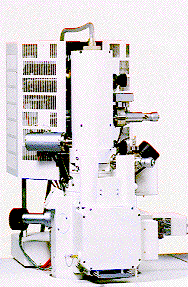
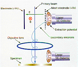
| A scanning electron microscope generates an image of a sample by bombarding it in vacuum with a well focussed electron beam and collecting the secondary electrons emitted by the sample. The top photograph shows the "head" unit of a Hitachi S-4700 SEM. The electron beam starts from a field emission tip at the top of the microscope and is focussed on the sample plane by a series of electron lenses. The beam is scanned over the sample in a raster pattern and the picture contrast comes from the change with scan location of the number of secondary electrons. To prevent sample charging and image degradation the specimen must frequently be coated by a thin carbon or gold film. The lower diagram shows the two detectors for this microscope. The lower detector is a conventional secondary electron detector, the top unit permits filtering the secondary electrons before they are detected for improved image quality. The resolution of the secondary electron image is as high as 2.5 nm at a primary beam energy of 1 kV. | ||||||||||
 |
||||||||||