Purkinje Neurons
Dmitry Sarkisov GS
Department of Physics
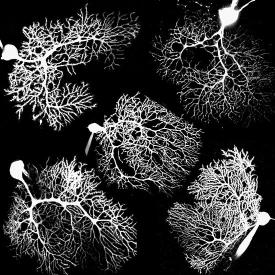
This is a composite image of five Purkinje neurons from the rat cerebellum, in the back of the brain. Each has been filled with fluorescent dye through a glass pipette, shown touching the cells. Images were taken on a two-photon microscope. Each Purkinje cell receives hundreds of thousands of inputs through its dendrite, the elaborate tree-like structure seen emerging from the cell body.
|