Research: Piezoelectric Microcantilevers
Christopher R. Martin
The focus of the piezoelectric microcantilevers effort is to produce sensors that can be readily utilized for a variety of applications. One such focus of the group is the development of biosensors capable of measuring concentrations of particular proteins in aqueous media [1-2]. These sensors could be injected into patients for in vivo sensing.
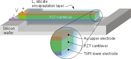
Schematic of a coated piezoelectric cantilever in use as a biological sensor.
Recent work by Christopher Martin has focused on the processing of micrometer-scale patterns of lead zirconate titanate (PZT, Pb(Zr,Ti)O3), a ferroelectric ceramic material with a high piezoelectric coefficient. The material is deposited from a sol-gel [3] using the soft lithography technique of micromolding in capillaries (MIMIC) on a variety of substrates to produce patterns as fine as 0.5 μm.[4-8] Soft lithography permits low cost fabrication of patterned thin films, imperative for the development of a commercial device; this approach is cheaper and simpler than the alternative of micromachining quartz.
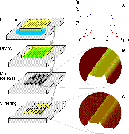
Schematic of MIMIC used to micropattern PZT thin films; insets show AFM images that depict the topographical deviation of the patterned features from the shape of the rectangular channels.
We recently completed a study to understand and control the topographical distortions from the shape of the original mold that results during the shrinkage of patterned thin film during drying and sintering.[9] These topographical defects limit the potential wide-spread application of MIMIC in microfabrication as they can hinder alignment and deposition of proceeding layers. The factors that contribute to this effect have been explained and modifications to the basic MIMIC technique have been developed to eliminate these distortions.
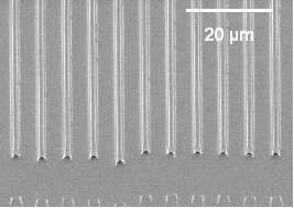
SEM image of PZT lines deposited on a silicon wafer by soft lithography
A concurrent effort is underway to understand the device physics of PZT cantilevers. For this work, millimeter- to centimeter-scale PZT pieces are mounted onto stainless steel foils and cut into cantilevers. Using an impedance analyzer, these cantilevers have been shown to be capable of measuring liquid viscosity and density.[10-11] The current focus is to compare the sensitivity of the microscale devices with millimeter-sized cantilevers prepared from commercial PZT films to validate the scaling relationships that have been derived in the literature. Further, we are comparing the cantilever design to the technique of Quartz Crystal Microbalance (QCM) to determine whether or not the cantilever design is indeed a better geometry for small mass detection.
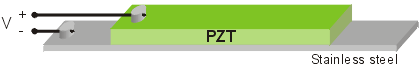
Schematic of millimeter-scale PZT cantilevers backed on stainless steel foil.
The ultimate aim of the project is to manufacture and demonstrate the functionality of a micrometer-sized PZT cantilever with electrodes. The successful application of this process will be the first demonstration of the use of soft lithography with silicon processing to produce a device as small as one micrometer in width.
References
1. J. Fritz, M.K. Baller, H.P. Lang, H. Rothuizen, P. Vettiger, E. Meyer, H.-J. Güntherodt, Ch. Gerber, J.K. Gimzewski, Science 288 (2000).
2. D.L. Polla, L.F. Francis, Annu. Rev. Mater. Sci. 28 (1998).
3. G. Yi, Z. Wu, M. Sayer, J. Appl. Phys. 64 (1988).
4. Y. Xia, G.M. Whitesides, Annu. Rev. Mater. Sci. 28 (1998).
5. E. Kim, Y. Xia, G.M. Whitesides, J. Am. Chem. Soc. 118 (1996).
6. W.S. Beh, Y. Xia, D. Qin, J. Mater. Res. 14 (1999).
7. S. Seraji, Y. Wu, N.E. Jewell-Larson, M.J. Forbess, S.J. Limmer, T.P. Chou, G. Cao, Adv. Mater. 12 (2000).
8. J.S. Vartuli, M Ozenbas, C.M. Chun, M. Trau, I.A. Aksay, J. Mater. Res. 18 [5] 1259-65 (2003).
9. C.R. Martin, I.A. Aksay, J. Phys. Chem. B 107 [18] 4261-68 (2003).
10. W. Y. Shih, X. Li, H. Gu, W.-H. Shih and I. A. Aksay, J. Appl. Phys. 89 [2] 1497-1505 (2001).
11. J. W. Yi, W. Y. Shih, W.-H. Shih, J. Appl. Phys. 91 [3] 1680-6 (2002).
For more information, please contact Chris Martin or use the links on the left to access additional information on the CML web site.
![]()
![]()
![]() © 2004 Princeton University, Ceramic Materials Laboratory. All Rights Reserved.
© 2004 Princeton University, Ceramic Materials Laboratory. All Rights Reserved.