Princeton's world-class microscopes are making a giant impact in the realm of the minuscule
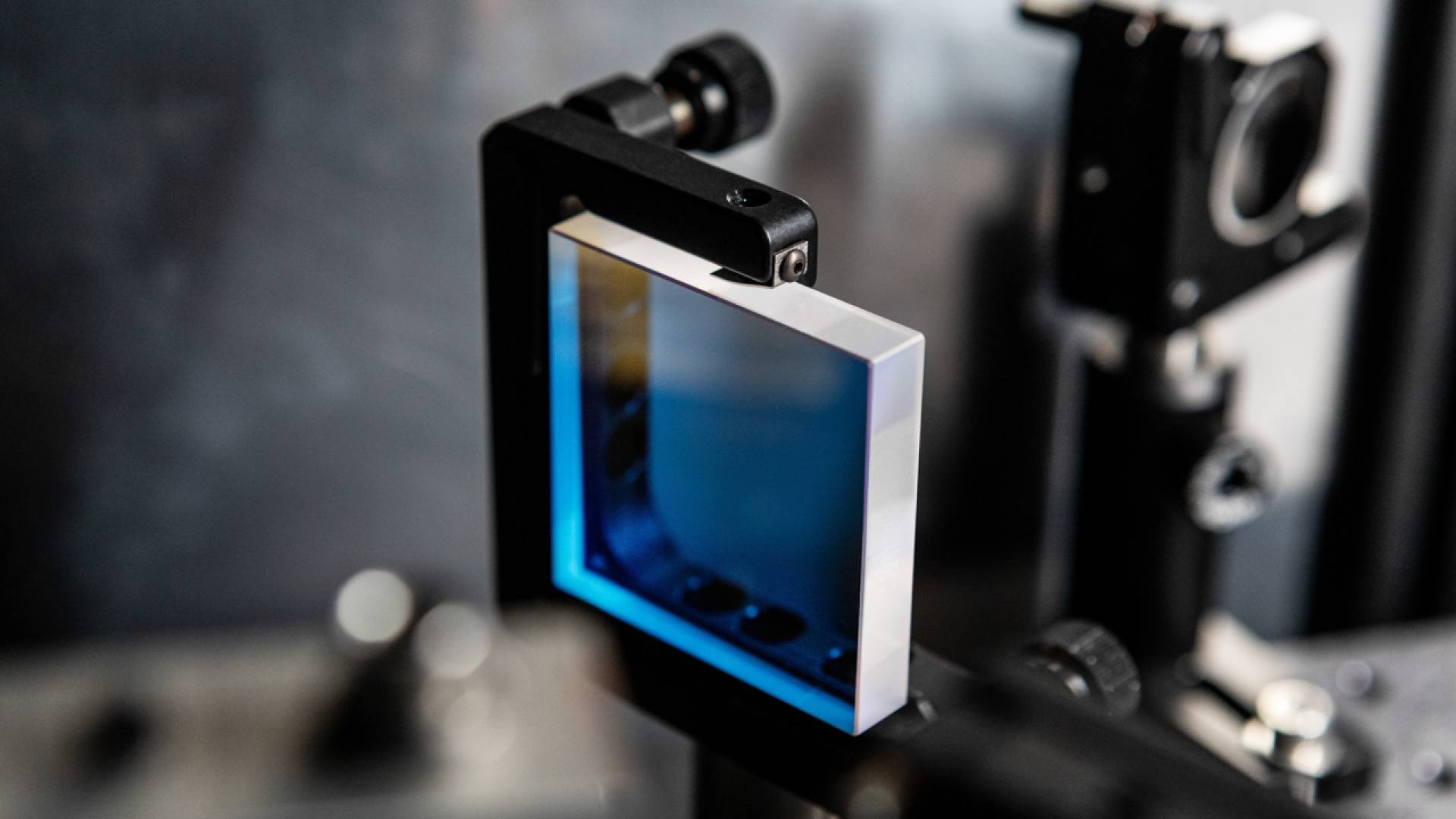
Princeton's microscopy tools range from this tiny near-infrared mirror, part of a light-bead microscope, to room-sized instruments with extraordinary sensitivity.
Princeton scientists are peering into the smallest corners of matter using an exceptional collection of sophisticated microscopes — some so big they fill a room. These remarkable instruments have established the University as a world leader in microscopy and led to countless discoveries.
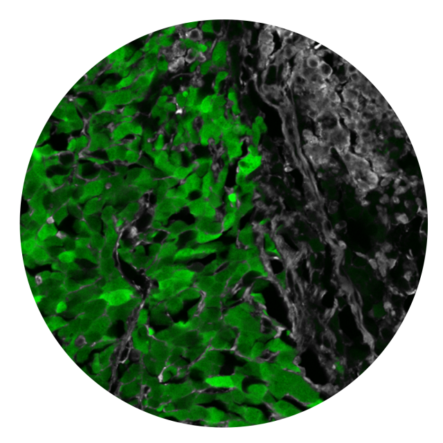
This fluorescent image from Yibin Kang's cancer research shows tumor cells (green) metastasizing into bone (black and gray). Visualizing the structure of the cells is a key to developing new treatments.
A way to stop cancer cells from metastasizing. New insight into Alzheimer’s. A revolutionary form of matter with 5-fold symmetry. Exquisitely precise quantum sensors built from diamonds.
“If you grab somebody on the street and say, ‘What do you care about?’, they’re not going to say ‘Microscopy!’,” said Cliff Brangwynne, a pioneer in the molecular forces behind neurodegenerative diseases and director of Princeton’s Omenn-Darling Bioengineering Institute.
“They’ll say, ‘I care about my health.’ ‘I care that I don’t lose my memories.’ ‘I care that the water’s clean.’ ‘I care that my phone works.’
“But all these things are dictated by microscopic processes that we cannot see with the naked eye,” said Brangwynne, who is also Princeton’s June K. Wu '92 Professor of Chemical and Biological Engineering. “Fundamentally, microscopy is a window into the underlying drivers of things that matter to everyone.”
A wonderland of world-class instruments
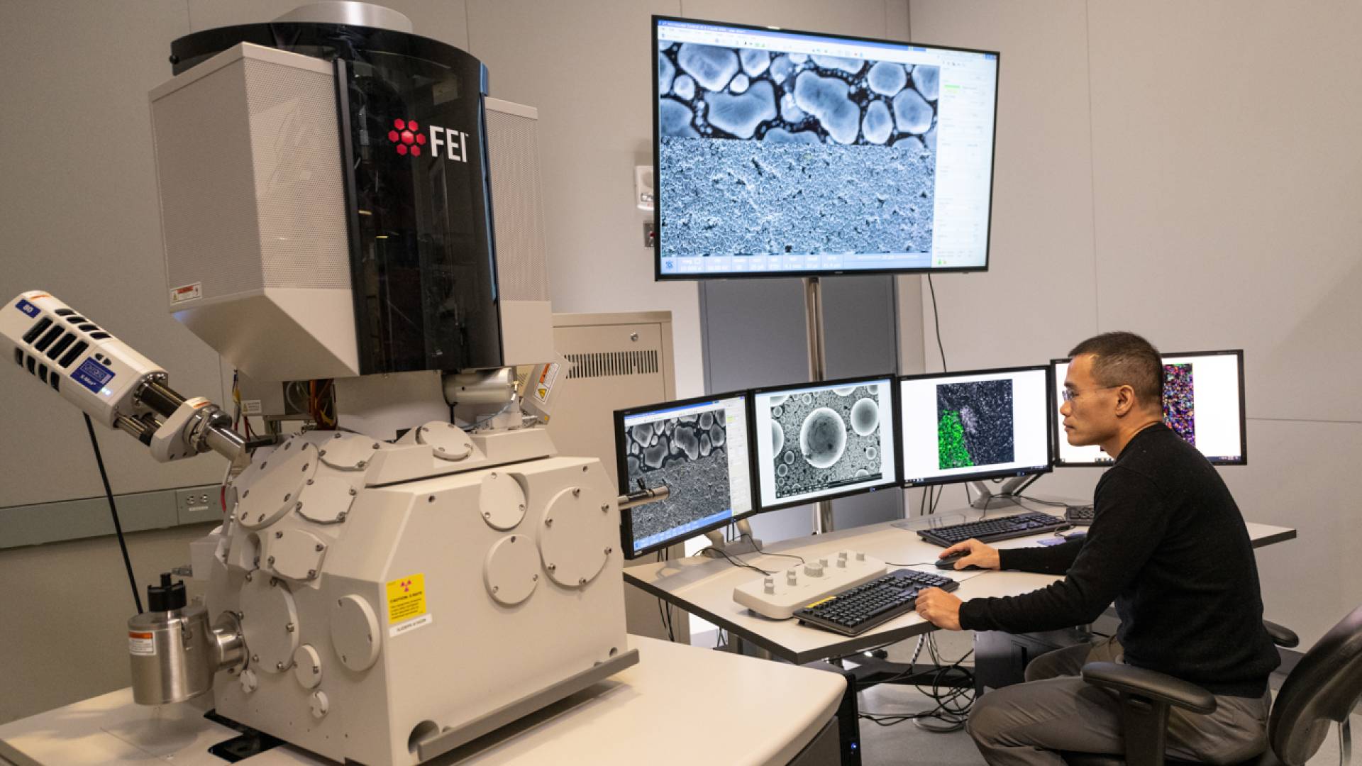
The University’s extraordinary microscopic and nanoscopic instruments include atomic force microscopes, transmission electron microscopes, X-ray diffraction and 3D X-ray microscopes, cryo-electron microscopes, ion and electron dual-beam scanning microscopes, and many others. Scientists and engineers use them to see inside human cells and map out the structures of matter, genes and proteins, building an understanding of microscopic processes that leads to new medicines, new tools and new materials.
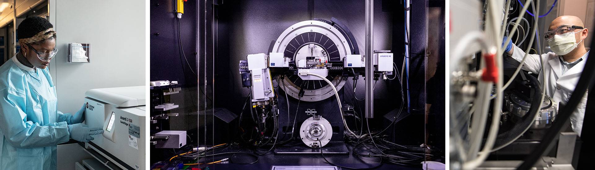
From left: Junior Jennifer Nwokeji prepares a sample in a centrifuge for a cryo-electron microscope; an instrument in Princeton's Imaging and Analysis Center awaits its next user; Paul Shao, senior imaging and analysis specialist at the IAC, adjusts a microscope.
Funding for these technological marvels comes from federal research grants, donor funding and the University endowment, which has helped purchase what Dean for Research Peter Schiffer calls “some of the best technical equipment in the world, in service of cutting-edge science and engineering research.”
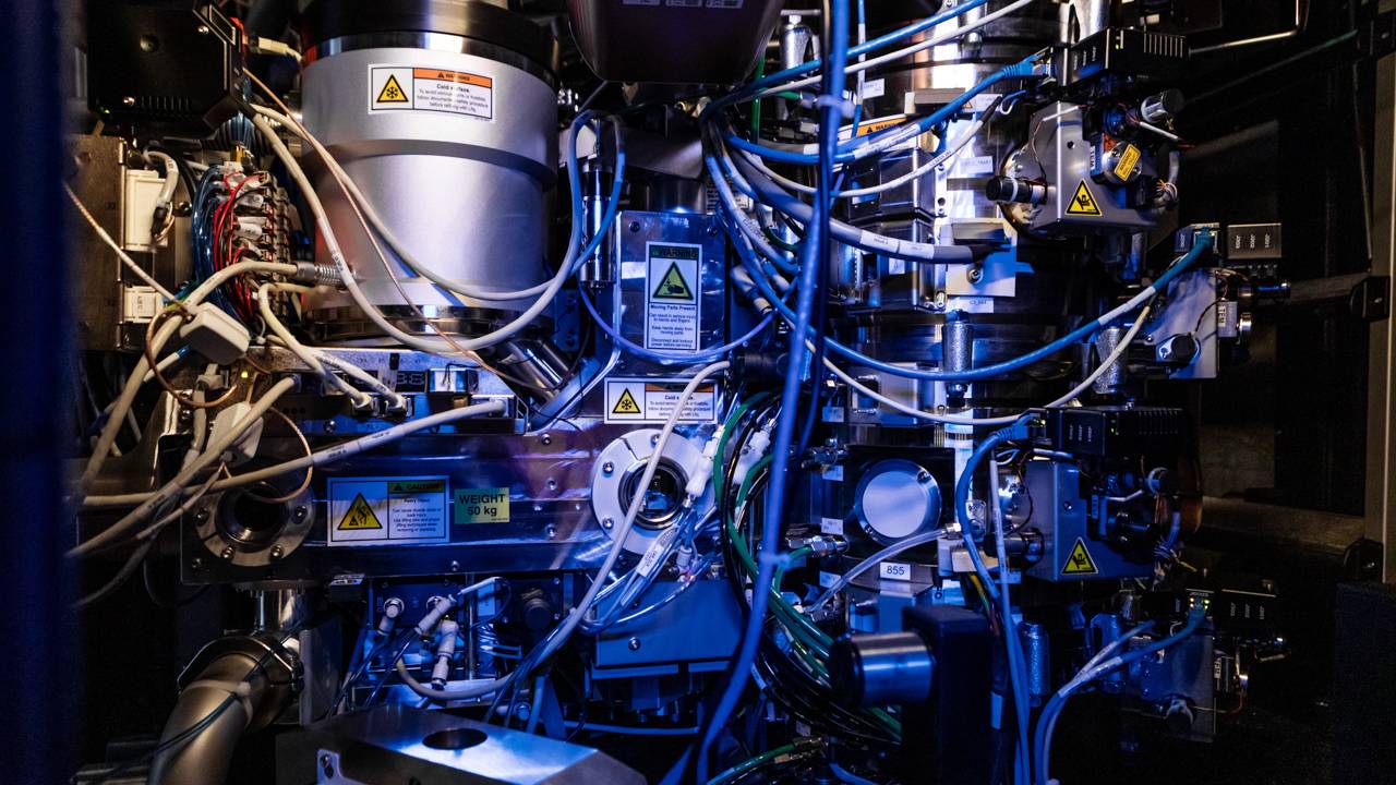
Many of the microscopes are housed together in the Imaging and Analysis Center (IAC), a wonderland of cutting-edge instrumentation available to all Princeton researchers, from undergraduates to senior scientists. Part of the Princeton Materials Institute, the IAC is located on the lowest level of the Andlinger Center for Energy and the Environment, where picture windows on both sides of a long hallway reveal instrument rooms containing microscopes that range in size from a toaster to an industrial freezer.
These exquisitely sensitive instruments need to be located underground, spiked into bedrock and isolated from even the smallest vibrations. Many of the microscopes require water chillers and noisy air quality handlers, all hidden behind sound-proofed walls. The entire facility is in encased in 3/8” thick aluminum to shield it from errant electromagnetic waves, and the heating and cooling systems are designed to move air in smooth sheets so as not to cause even an atom-sized jiggle.
While many of the University’s largest instruments are in the IAC, many more microscopes are located in shared facilities or in individual labs around campus. One of the newest, a multi-million-dollar instrument being built by Herschel Rabitz, the Charles Phelps Smyth ’16 *17 Professor of Chemistry, and Martin Jonikas, associate professor of molecular biology, will be housed in Frick Chemistry Laboratory. This recently funded project aims to develop an instrument that provides unprecedented views of chemicals and small molecules at nanometer resolution.
“The ready availability of these tools enables our researchers to make advances that lead their fields,” said Schiffer, a professor of physics. “Furthermore, world-class facilities attract other top scholars, who in turn share their expertise and insights, leading to more progress, which attracts more excellent researchers, creating a virtuous cycle.”
These multi-user facilities also serve as a focal point of interaction with industry. Companies with research questions pay to use many of the facilities, which speeds innovation while injecting ideas from real-world uses into teaching and research.
“Microscopy is an engine to all kinds of research, from environmental science to bioengineering and quantum physics,” said Nan Yao, the IAC director and the inaugural professor of the practice in the Princeton Materials Institute. “No matter what kind of materials we deal with — polymers, ceramics, glass, graphene, quantum dots, lithium metal oxide, or living cells — we need to see it to analyze it.”
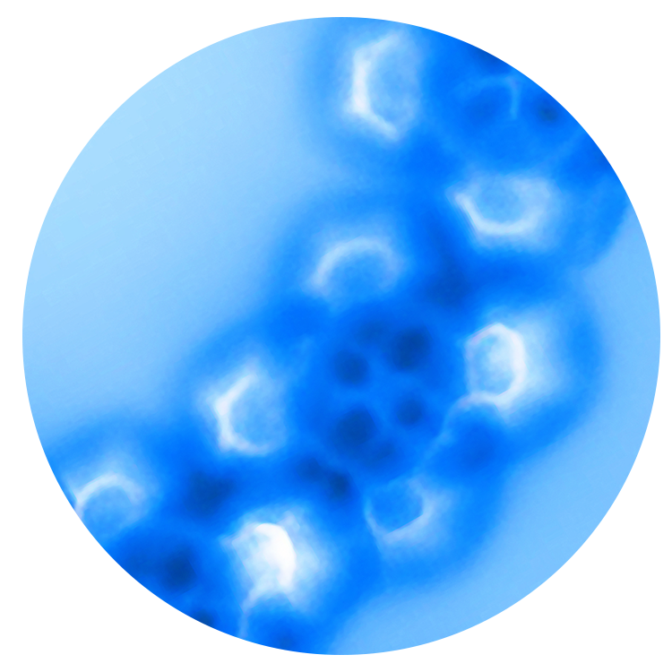
An atomic force microscope at the IAC showed the atomic structure of cobalt and iron, elements that differ only by one proton and one electron. The iron atom has a squarish shape, while cobalt is brighter with more distinct lobes in its electron orbitals.
Microscopic discovery at Princeton
Princeton researchers and engineers are peering into living cells, newly discovered materials, and many more things at impossibly small scales.
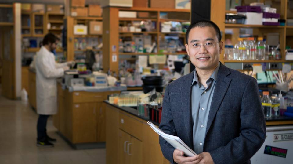
- Cancer biologist Yibin Kang found a way to stop cancer cells from metastasizing after X-ray crystallography revealed the structures of two intertwined proteins.
- Nan Yao and physicist Paul Steinhardt used two IAC instruments to confirm the existence of naturally occurring “quasicrystals,” a material with 5-fold symmetry that was believed for centuries to be impossible.
- Engineer Lynn Loo used IAC tools to develop a new material that, when layered only a few atoms thick between perovskite and electronics, makes perovskite solar cells commercially viable.
- Evolutionary biologist Lindy McBride uses a custom-designed microscope to identify how mosquitoes target humans.
- Quantum physicist Nathalie de Leon is engineering dual-nitrogen-vacancy quantum sensors from diamonds with the help of IAC tools.
- Biophysicist Joshua Shaevitz and neuroscientist Andrew Leifer designed and built a groundbreaking custom microscope to image the whole brain of a living, moving worm.
- Environmental engineer Jason Ren used IAC spectroscopy and electron microscopy in developing his radical new approach to extracting lithium, a vital component of the batteries at the heart of a clean energy future.
- Yao and collaborators across the University and the Princeton Plasma Physics Laboratory used the IAC’s atomic force microscope to capture the first images of the electron orbitals (shown here) within individual atoms.
Yao has run the IAC since its inception and helped it grow from housing a single instrument to establishing Princeton as a world leader in microscopy. “Of all the top-notch papers published by Princeton authors in Science, Nature and similarly high-impact journals, fully half are associated with the Imaging and Analysis Center,” he said. “Either an author was our user or they used imaging data collected here. This facility is a very important investment for the University.”
In recognition of the IAC’s unique capabilities, Nature co-leads a “Frontiers in Electron Microscopy for Physical and Life Sciences” conference with Princeton every two years.
The conference brings together top microscopists from around the world to share discoveries, approaches and insights with Princeton’s own microscopy community — the hundreds of undergraduates, graduate students, postdoctoral researchers, staff researchers and faculty members whose advances depend on observing the smallest realms.
A community of curiosity-driven researchers
It is the community of curious scientists, as much as the technology, that distinguishes microscopy at Princeton.
“The beauty of Princeton is that we combine both physical sciences and life sciences together in the IAC, so researchers come here, talk to each other, and start interdisciplinary collaborations,” Yao said.
Additionally, “here at Princeton, the people who do the analysis talk with the people who think about instrument design and the people who write the algorithms. That sets us apart,” said Joshua Shaevitz, a professor of physics and the Lewis-Sigler Institute for Integrative Genomics, the director of the graduate program in biophysics, and a longtime leader in microscopy at Princeton. “Our goal is not just to push the resolution smaller and smaller, it’s about driving discovery.”
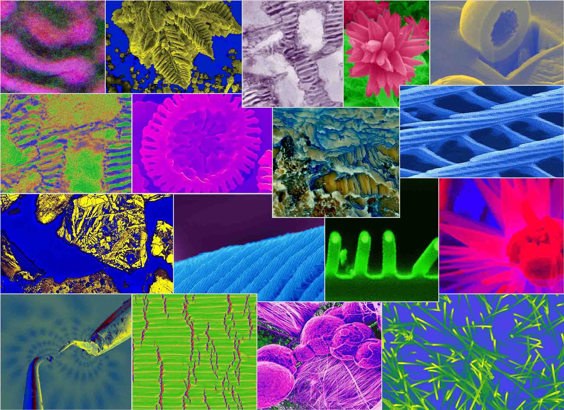
These images were all generated from instruments in Princeton's Imaging and Analysis Center (IAC), which is run by the Princeton Materials Institute and located in bottom level of the Andlinger Center for Energy and the Environment.
For his work on metastatic breast cancer, for example, Yibin Kang connected with colleagues who used X-rays to visualize metadherin, a protein that Kang knew was vital to the spread of deadly cancers. The imaging revealed that the protein has two finger-like projections that stick into two pockets on another protein, SND1. Kang’s team had had little success disabling metadherin itself, but after seeing the interconnection, they used high-throughput screening assays to identify a molecule that fills one of SND1’s pockets. They modified that molecule, changing it into a compound that prevents the proteins from intertwining.
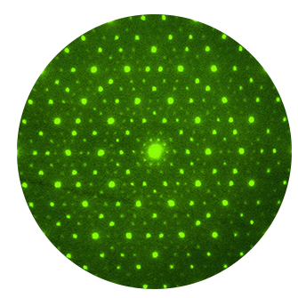
This pattern confirmed the existence of a decagonal quasicrystal, a material believed for centuries to be impossible.
“That’s the beauty of biology,” said Kang, who is the Warner-Lambert/Parke-Davis Professor of Molecular Biology at Princeton University, a founding member of the Ludwig Institute for Cancer Research Princeton Branch, and an associate director of Rutgers Cancer Institute of New Jersey. “Crystal structures give you a crystal-clear vision about how, at the atomic level, biology works. Seeing the crystal structure gave us the insight to develop the compound, and then we imaged it again and saw that it binds to the pocket exactly as we had hoped and predicted it would do.” That compound is now making its way through the FDA’s drug development process.
Princeton microscopes and teamwork also played a key role in a revolutionary contribution to materials science: discovering a natural quasicrystal. For centuries, crystallographers had insisted that crystals could only have 3-fold, 4-fold, or 6-fold symmetry, never 5-fold or 7-fold.
But Yao and physicist Paul Steinhardt, Princeton’s Albert Einstein Professor in Science, believed that 5-fold symmetry must exist in the natural world, since other scientists had artificially created a quasicrystal in a lab.
Their search for a naturally occurring quasicrystal took more than a decade, but Yao and Steinhardt eventually found the world’s first, and then second, naturally occurring quasicrystals. “Without electron imaging and diffraction, this would have been impossible,” Yao said. “The crystal sample we had was so small, no other technique could have detected its detailed structures.”
The IAC eagerly welcomes undergraduates into the community and supports their scientific inquiry. “Within two weeks, I went from not knowing much about what an electron microscope even is to using one for my own data acquisition,” said sophomore Maria Karakousis.
Junior Jennifer Nwokeji is using one of the University’s most sophisticated microscopes, a cryogenic electron microscope (cryo-EM), to try to decode how a protein is burrowing through the membrane wall of a human cell. She hopes to find a cure or treatment for malaria, a disease that still ravages her parents’ home country of Nigeria.
The microscope is as big as a walk-in closet, and one of two cryo-EMs in the IAC. It offers an unparalleled view inside cells and will help her build a 3-D model of the attacking protein. “For proteins, structure dictates function,” Nwokeji said. “Once we have the structure, future research can see what it binds to, what it does and ultimately, how to design a drug to stop it.”
Bonnie Bassler, chair of the Department of Molecular Biology, was instrumental in bringing the cryo-electron microscopes to Princeton. “For half a century, X-ray crystallography reigned supreme as the best tool available to peer into the world of the small,” said Bassler, who is also Princeton’s Squibb Professor in Molecular Biology and a Howard Hughes Medical Institute Investigator. “It’s what Rosalind Franklin used to discover the structure of DNA. It is great for seeing an individual protein, or even maybe two. But cryo-EM shows us whole enormous machines inside a cell.”
A cryo-EM costs about $10 million.
In 2016 Princeton invested endowment funds to buy one, even though no cryo-EM experts were on the faculty at the time.
“We might have bought it and been the proud owners of a $10 million paperweight,” said molecular biologist Bonnie Bassler. “But Princeton took that bet.”
The University worked closely with donor Gerhard Andlinger to design the IAC to house the delicate instrument, which needs to be grounded in bedrock.
The cryo-EM bet paid off, as one after another top cryo-EM expert joined the faculty and trained colleagues.
As demand has grown, the University has now bought a second cryo-EM. The IAC houses these and 31 other leading-edge microscopes.
Building bespoke microscopes
In addition to the room-sized microscopes available in the IAC, many Princeton researchers and engineers design instruments targeted to their specific questions, which are then housed in their own labs.
“There are so many different flavors of microscopy,” said Brangwynne. “There’s huge difference whether you’re imaging materials, electronic devices or living systems, or whether you’re using an instrument to observe or to manipulate materials. Do you need to see a single cell? Is it a small cell or a big cell? Or is it the 10 million cells making up lung tissue? Microscopy comes in thousands of different flavors, and each has specific needs and preferences.”
Stéphan Thiberge, director of microscopy facilities at the Bezos Center for Neural Circuit Dynamics at the Princeton Neuroscience Institute, is like a modern-day clockmaker, assembling novel microscopes with tiny screwdrivers — with help from computer-assisted design software and the physics department’s machine shop.
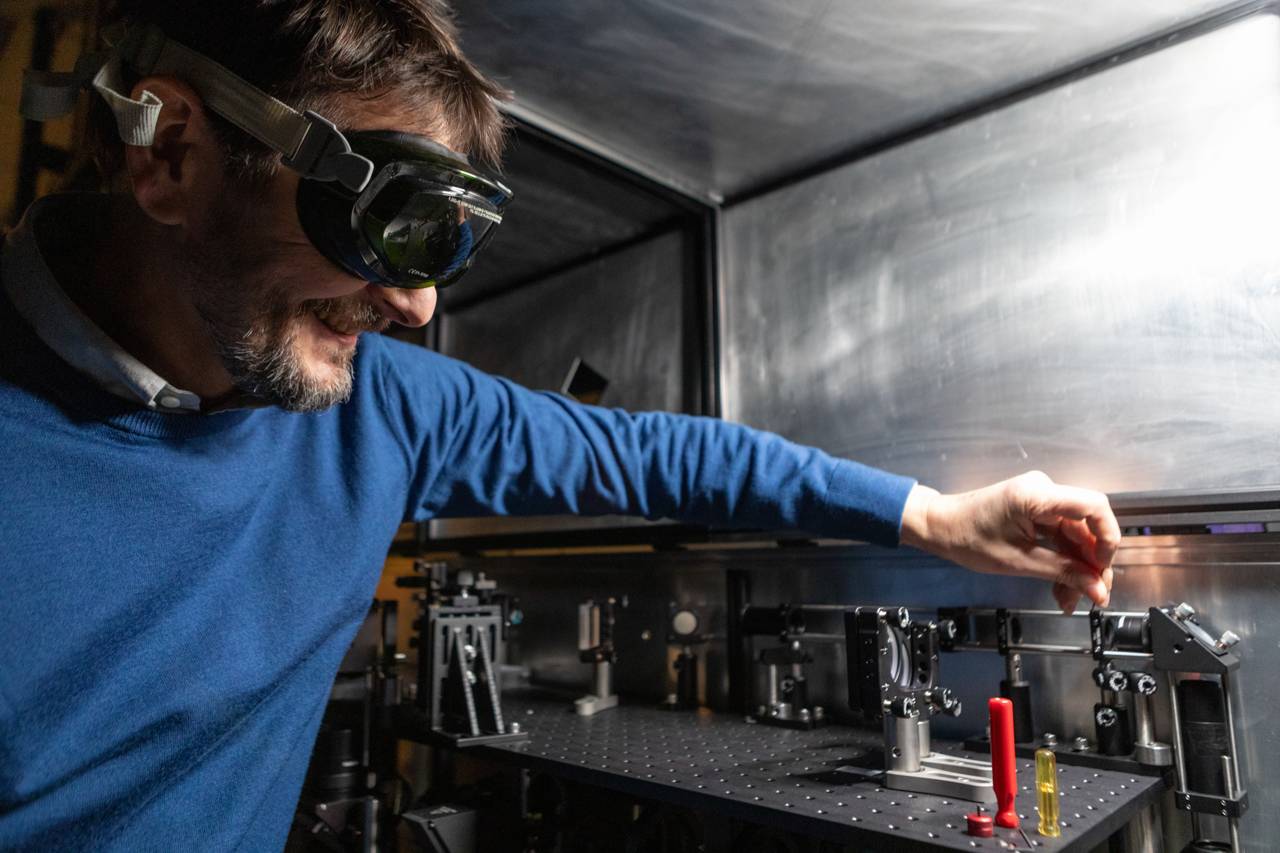
Microscope designer Stéphan Thiberge secures a lens in place in the optical path of a microscope.
Designing the instrument makes all the difference, said mosquito researcher Lindy McBride, an associate professor of ecology and evolutionary biology (EEB) and neuroscience. Key to her team’s discovery of how mosquitoes target humans, she said, was an instrument built by Thiberge to image the neurons inside a mosquito’s brain. “We could walk over to the Bezos Center and say we want to image this, at this resolution, with this orientation, and a few months later, the microscope was built,” she said. “We could have bought an off-the-shelf microscope, but it would have been so much slower and so much less powerful.”
For her work exploring bacterial communication, Bassler uses a microscope custom-built for her lab. “Bacteria are really tiny,” she said. “It's hard to image even individual bacteria. It's really hard to image single-gene mRNA transcripts inside bacteria. They’re smaller than small.”
Seeding the world with microscopists
Several scientists noted the importance of training undergraduates and graduate students to use these instruments so that they are prepared to lead their own research labs and accelerate the rate of discovery.
“One of the beauties of the IAC is that this is not just a research center, but also an education center,” Yao said. Yao teaches two award-winning microscopy classes for undergraduate and graduate students. In addition, he and his team lead a two- to four-hour workshop every month on various microscopy techniques on all 33 instruments. Thousands of students have learned and practiced microscopy in the IAC.
Princeton undergraduates who take advantage of the IAC training and resources go on to graduate school with a clear edge, he said. At most peer universities, he said, only graduate students and postdocs have access to the most cutting-edge tools.
Princeton’s microscopes and its commitment to making them accessible both played a role in recruiting several recent hires in molecular biology and engineering and applied sciences, including two who were renowned cryo-electron microscopists before coming to the University.
A.J. te Velthuis, an assistant professor of molecular biology and a leading virologist who studies coronaviruses and flu viruses, said he was drawn to Princeton because of its community of instrument users, who meet regularly for scientific discussion and to share insights and ideas. “During my interview, I spoke with students and members of the faculty,” he said. “Getting to work with them and find new ways to look at viruses is really inspiring.”
John Jimah, an assistant professor of molecular biology who studies malaria and related parasites and has contributed to the development of a transmission- and infection-blocking malaria vaccine candidate, said, “One reason I chose to come here was that I could see that the environment was very interested in training and mentoring the next generation of scientists.”
Jimah, who is Nwokeji’s advisor, continued: “Microscopy is a fast-moving field, and Princeton continues to invest in it. To keep making world-class contributions to research, we and our trainees need to use world-class equipment, both for discoveries and to bring those discoveries out to the wider world.”
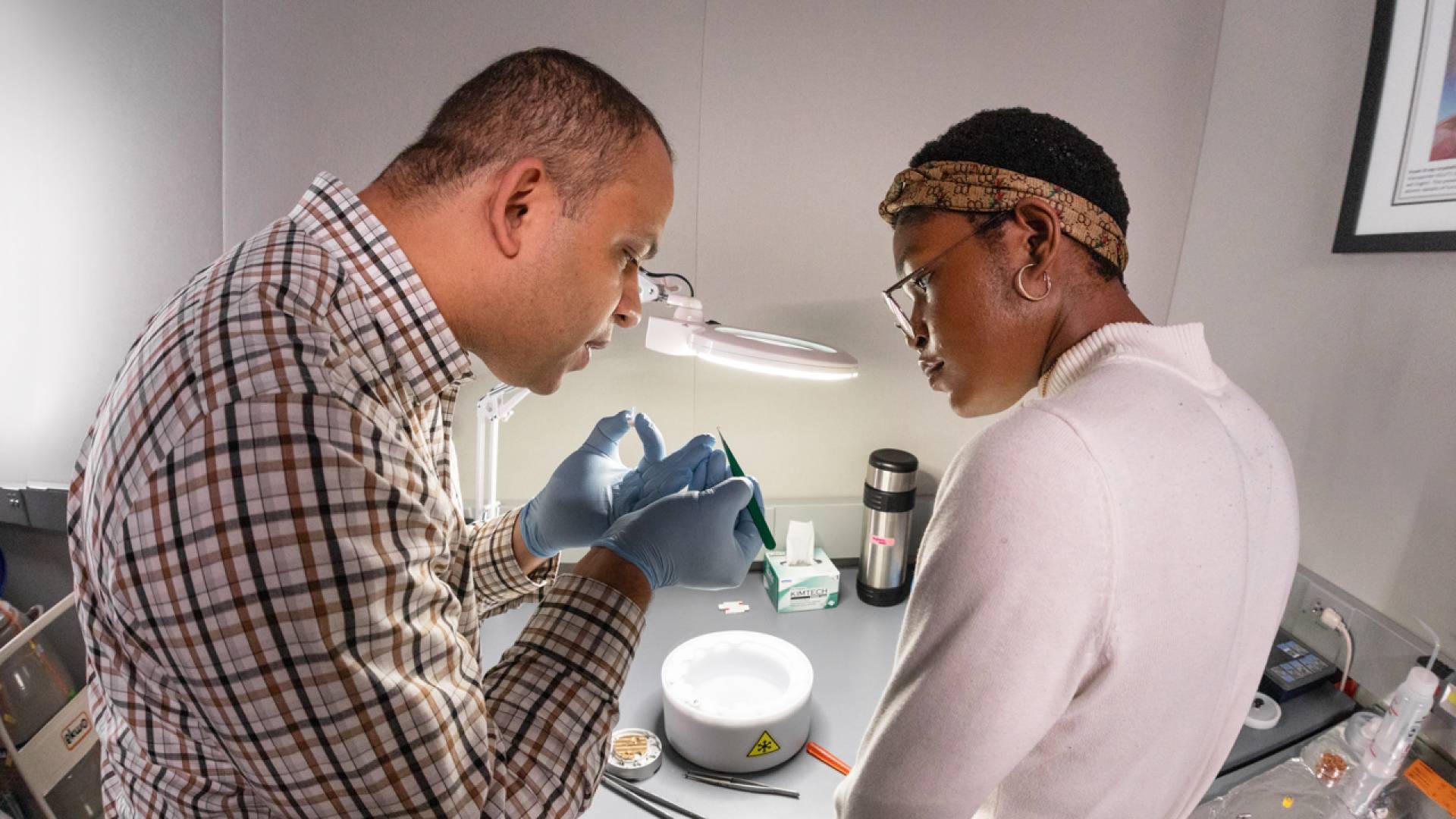
Biologist John Jimah and junior Jennifer Nwokeji prepare a sample for the cryo-EM.
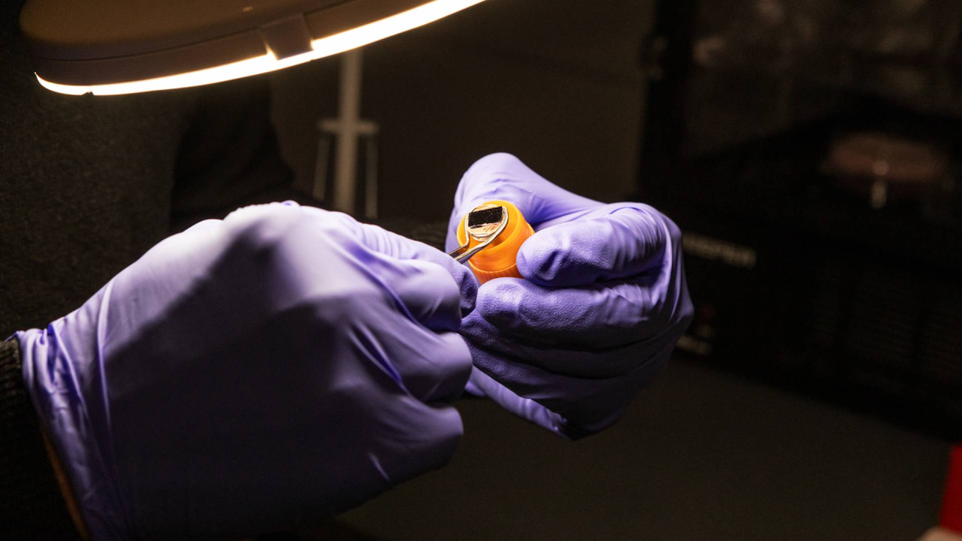
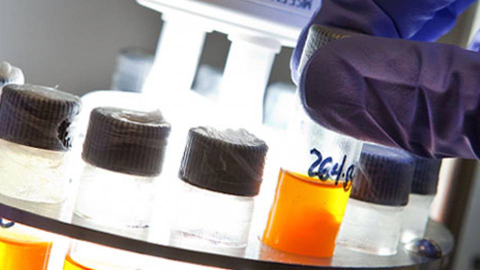
A researcher prepares a sample for electronic microscopy imaging.
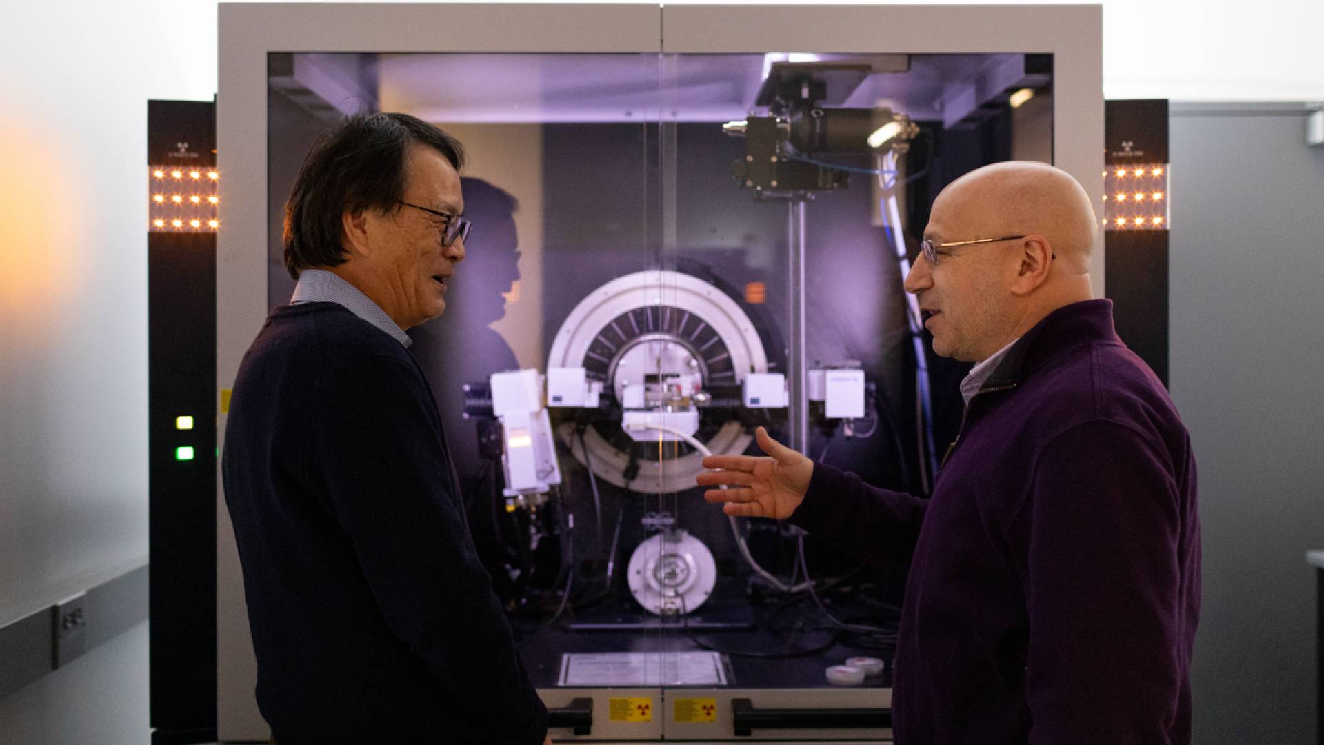
IAC Director Nan Yao and Howard Stone, Princeton's Donald R. Dixon '69 and Elizabeth W. Dixon Professor of Mechanical and Aerospace Engineering, talk in front of one of the IAC instruments.
Cancer biologist Yibin Kang looks at samples with a scanning electron microscope.
Microbiologist Bonnie Bassler talks with Paul Shao at the IAC.
Federal grant support for the Princeton researchers included in this story comes from agencies including the National Science Foundation, the National Institutes of Health, the National Cancer Institute, the Department of Energy and the Department of Defense.