Decaying Oak Leaf II(b): Attrition
Alessandra Leri GS
Departments of Chemistry and Geosciences
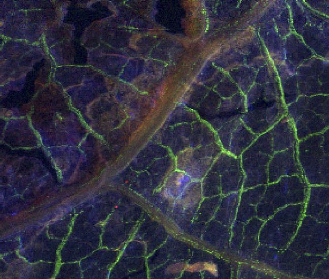
X-ray fluorescence microscopy allows geochemists to map the distributions of many different elements simultaneously in chemically heterogeneous natural samples. This is an X-ray fluorescence micrograph of a 5 x 6 mm section of a degraded white oak leaf taken from the forest floor in the Pine Barrens, New Jersey. Calcium is highlighted in green, manganese in red, and chlorine in blue. (Lighter color corresponds to more intense fluorescence, i.e. greater concentration.) Manganese appears concentrated in the large veins and in a more diffuse background concentration throughout the leaf tissue. Calcium is localized primarily in the smaller leaf veins. Chlorine occurs in particularly high concentration in the center of the image, near the delta formed by the leaf veins. Optical microscopy revealed that the area of high chlorine concentration coincided with the presence of a mass of fungi colonizing the leaf surface, implicating the fungi as important players in the natural chlorination of decaying plant material. Special thanks to Matthew Marcus, Lawrence Berkeley National Laboratory, Advanced Light Source, Berkeley, California.
|