Three projects with the potential to open new avenues in science and engineering — mapping the body’s “electrome,” miniaturizing imagers to monitor the environment and human health, and accelerating gene editing for the treatment of disease — have been selected for funding through Princeton’s Eric and Wendy Schmidt Transformative Technology Fund.
“These are early-stage projects with tremendous potential to enable scientific discoveries and technological innovations that can benefit society at large,” said Dean for Research Pablo Debenedetti, the Class of 1950 Professor in Engineering and Applied Science and professor of chemical and biological engineering. “These projects have transformative potential but are so groundbreaking that they carry some risk, and we are grateful to Eric and Wendy Schmidt for their generosity and their vision in recognizing the importance of funding this stage of research.”
The fund was created in 2009 through a gift from Eric and Wendy Schmidt. Eric Schmidt is the former Chief Executive Officer of Google and former Executive Chairman of Alphabet Inc., Google’s parent company. Wendy Schmidt is a businesswoman and philanthropist. Eric Schmidt earned his bachelor’s degree in electrical engineering from Princeton in 1976 and served as a Princeton Trustee from 2004 to 2008.
The three projects were selected based on their capacity for broad and transformative impact:
Mapping the electrome

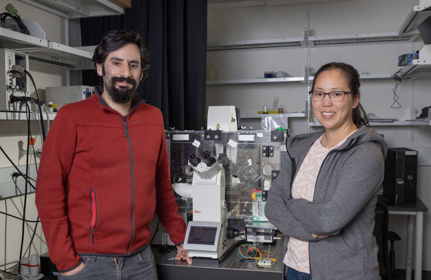
Daniel J. Cohen and Michelle Chan
- Daniel J. Cohen, assistant professor of mechanical and aerospace engineering
- Michelle Chan, assistant professor of molecular biology and genomics
Electricity is foundational to life, from the electrical regulation of our heartbeats to natural electric currents that help our injuries heal. Although clinicians have had some success using electrical stimulation to treat brain tumors and improve organ transplantation, researchers lack a clear mechanistic understanding of how electrical inputs translate to the body’s responses.
To enhance this understanding, this project aims to create comprehensive maps of the relationships between electrical inputs and resulting cellular behaviors. The team will create a new, high-throughput platform for conducting experiments to determine how electrical stimulation leads to associated responses, from the genes that become activated to behaviors such as cell migration and proliferation.
These maps of living organisms’ electrical landscape, or electrome, will form an atlas that researchers can use to predict how applying electrical stimulation can cause a corresponding change in a given cell type. The team brings together expertise in bioelectric interfaces and genomics to answer questions such as: What is the range of biological responses that electrical stimulation can target? Which types of electrical inputs give the optimal biological response? The answers could provide fresh perspectives in the treatment of a range of diseases.
Shrinking imaging to the atomic level

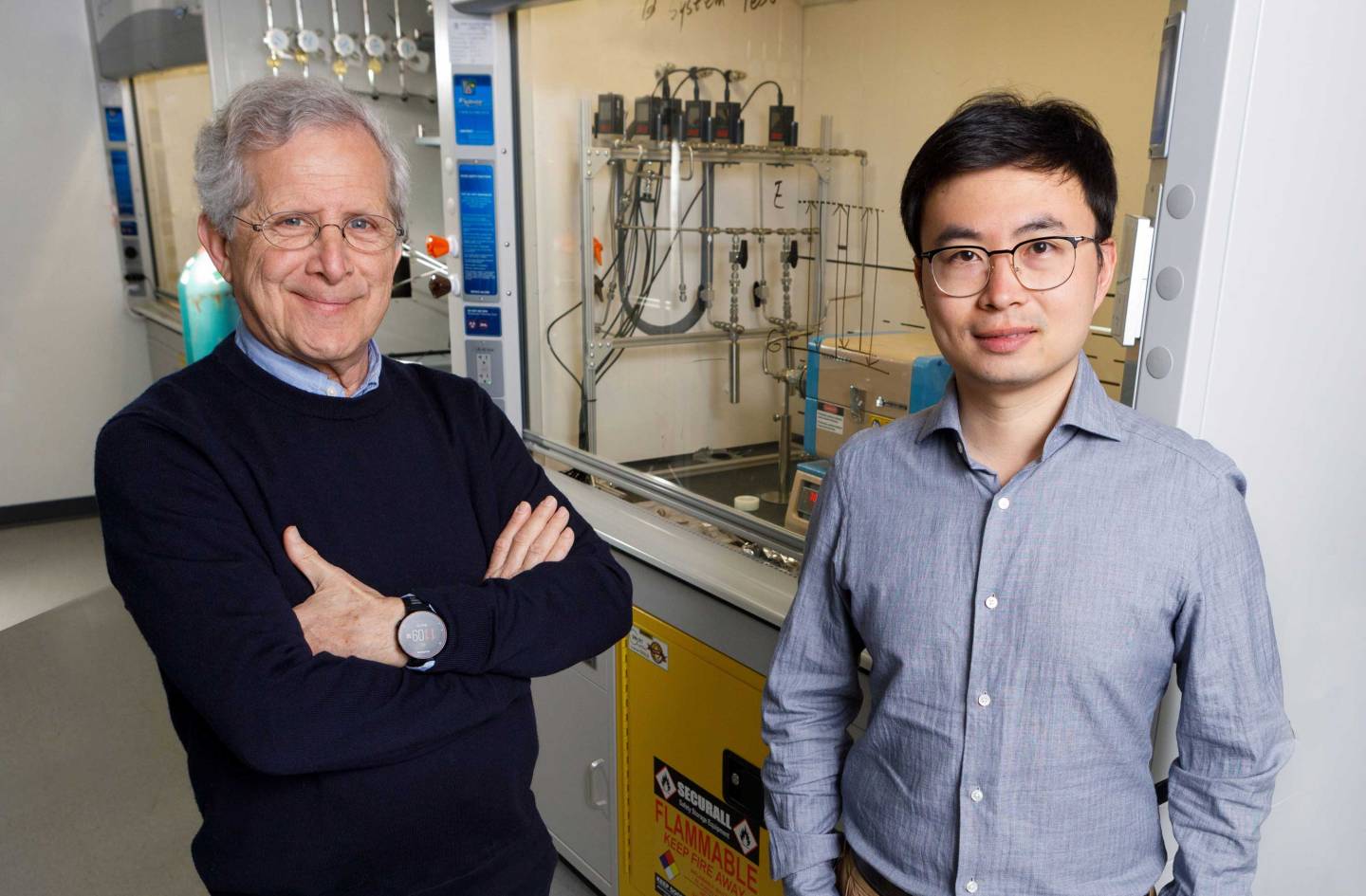
Antoine Kahn and Saien Xie
- Saien Xie, assistant professor of electrical and computer engineering and the Princeton Materials Institute
- Antoine Kahn, vice dean of the School of Engineering and Applied Science, the Stephen C. Macaleer '63 Professor in Engineering and Applied Science, and professor of electrical and computer engineering
Many areas of health and science involve evaluating the properties of a chemical or other object by measuring the range of wavelengths of light, or spectra, emitted by the substance. Spectrometers, devices that measure these emissions, are large and bulky because they first must run the light through a prism or filter that separates light into its different wavelength components prior to detection by silicon-based detectors, which alone cannot distinguish between light of different colors.
By harnessing new materials to build the detector, this project aims to build the world’s first atomically thin and lightweight imager capable of measuring the intensity of light at distinct wavelengths, for applications such as medical imaging, precision agriculture, environmental monitoring and self-driving vehicles. The small footprint and miniscule weight mean that the imager can be implanted in the body, used on wearable medical monitors, or mounted to a drone for aerial imaging.
The team, led by an expert in materials synthesis and a leader in optical, electronic and nanoscale spectroscopies, will create a new type of detector based on a class of two-dimensional materials known as transition-metal dichalcogenides. These materials can detect light of individual wavelengths, eliminating the need for prisms and filters and opening opportunities for creating novel imaging devices for a range of applications.
Expanding the capabilities of gene editing

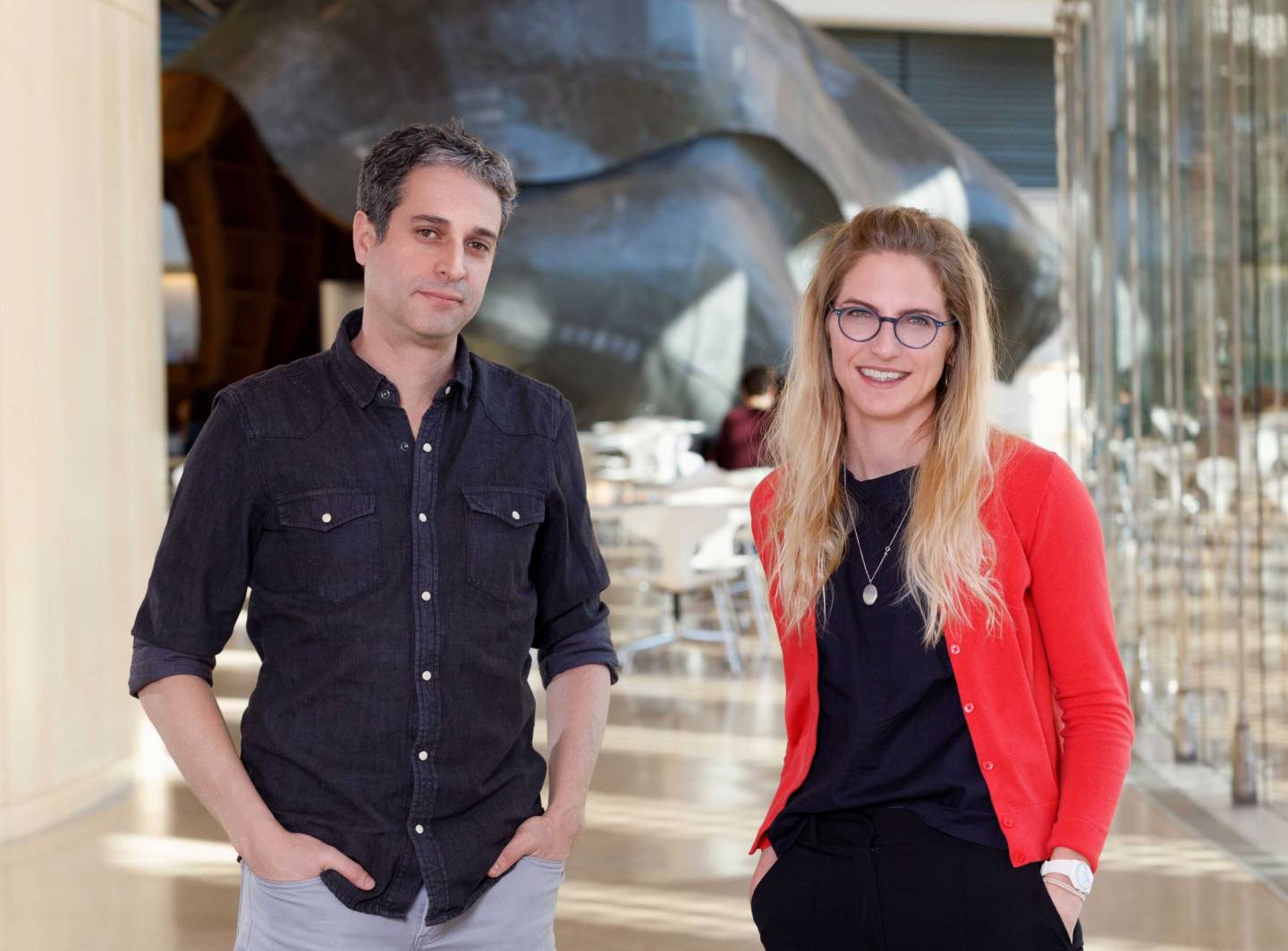
Ricardo Mallarino and Fenna Krienen
- Fenna Krienen, assistant professor of neuroscience
- Ricardo Mallarino, assistant professor of molecular biology
Our understanding of the human body and our ability to treat diseases rests in large part on research conducted in just a few model organisms, such as the laboratory mouse. To expand the repertoire of gene-editing approaches to additional mammalian species, this project will develop minimally invasive technologies that enable the addition or deletion of large DNA fragments and the ability to “write” new genetic information into a specific DNA site.
Unlike techniques commonly used in lab mice, this new approach relies on a strategy that delivers gene-editing reagents directly rather than removing and then reimplanting embryos. The ability to edit genomes with precision and ease promises to unlock the capability to study the animal model most relevant to the human biological process being studied, benefitting drug discovery as well as research on neurobiology, behavior, physiology and many other areas.
The team will include key technical expertise from Sha Li, a research associate in the Mallarino lab in a collaboration across neuroscience and molecular biology.


